Why Ultrasound
Thoracic Ultrasound
An echocardiogram (ECHO) is an ultrasound study of the heart. It allows us to see chamber size, valve structure, valve function, blood flow and velocity, and to evaluate the soft tissue/muscle completely.
In addition to an ECHO, an ultrasound of the mediastinum, lungs, or pleural space can also be performed to evaluate for lymphadenopathy, masses, fluid, etc.


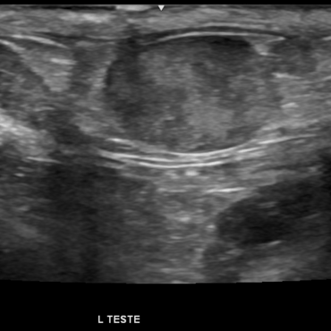
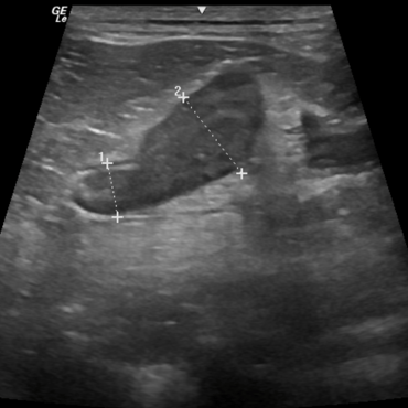
Soft Tissue Ultrasound
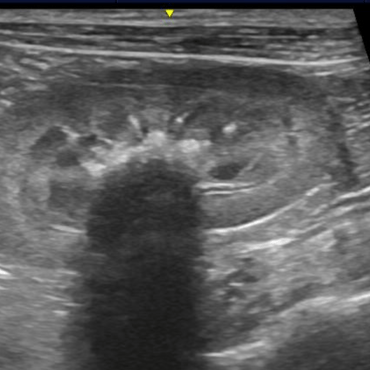
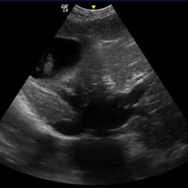
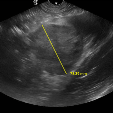
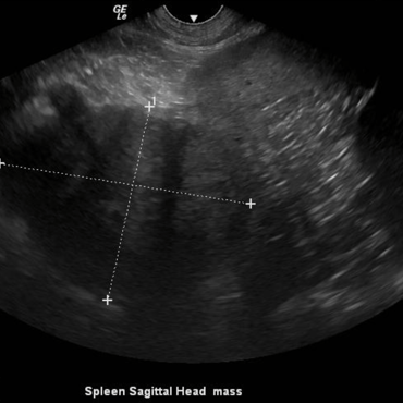
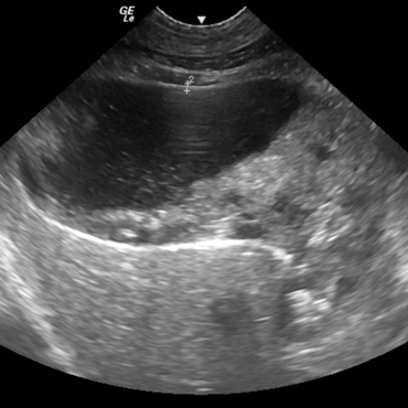
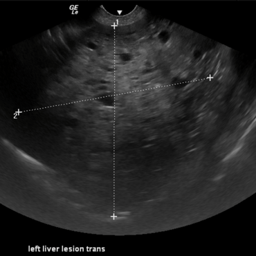
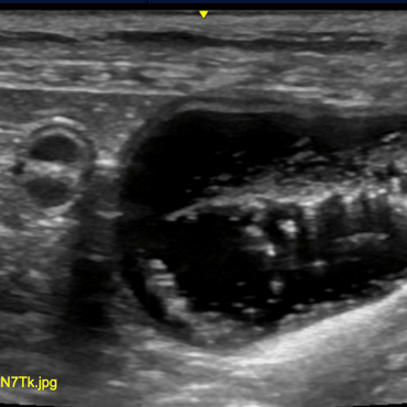
Ultrasound of the abdomen helps us to see the tissue structure of the various organs, vessels, and lymph nodes throughout the abdomen. This is particularly helpful when we are evaluating for masses, thickened luminal walls, thickened organs, cyst vs. mass, small sediment and stones that a regular radiograph cannot pick up on.
Liver shunts are not always visible/easy to determine via ultrasound. CT with contrast is the preferred method of identifying these structures.
Ultrasound Limitations
Ultrasound is my favorite imagine modality because it can help us distinguish so much that radiographs alone cannot. That being said, it still has its limitations. The most important one being that the ultrasound waves cannot penetrate bone, and they cannot penetrate air. Any gas within the GI tract can mar the structures beneath it and make it way more challenging to identify important structures and evaluate them. For this reason, sedation is always recommended to help minimize this issue and help pets be relaxed for their exams.
Fetchin' Veterinary Services
Bringing your clinic the help it needs.
Contact
info@fetchinvs.com
615-660-7334
© 2025. All rights reserved.